GE Logiq 7 Ultrasound
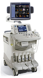
At the leading edge of healthcareThe LOGIQ 7 system provides a full range of clinical applications – including abdominal, small parts, surgery, vascular and cardiac imaging – and the power of GE's patented TruScan architecture.
Below is a generalized description of this systems technology, specifications, features and options. The below may not reflect the features and options available on units in our inventory.
|
- Technology
- Specifications
- Measurements & Reporting
- Transducers
Image quality is the cornerstone of the LOGIQ 7. GE has developed advanced technology that gives you improved resolution and superior image quality, for enhanced diagnostic capabilities that help you see more than ever before. GE's matrix technology diminishes the compromises between penetration and resolution.
Clear flow visualization |
|
| B-Flow, a unique GE technology, displays true hemodynamics and provides direct visualization of blood reflectors by magnifying those blood reflectors 30dB (1000 times). A significant advantage for vascular studies, it eliminates color flow over-write, frame-rate impact, and offers less angle dependence. |
|
CrossXBeam™ with Color Doppler, LOGIQView and Virtual Convex |
|
| A new Digital Compounding algorithm extends CrossXBeam™ advantages to Color Doppler examinations and Extended Field of View enabling you to get an unmatched image clarity and contrast resolution in any working modality. |
|
| Fewer speckles, more definition Speckle Reduction Imaging (SRI) heightens your visibility through improved contrast resolution. SRI is a new adaptive, real-time software algorithm that suppresses speckle artifact while maintaining true tissue architecture. |
|
Uncompromised penetration and resolution |
|
| Matrix array transducers with multiple rows of elements help you achieve uniform resolution throughout the field of view, which reduces volume averaging and improves overall image uniformity in both near and far fields. GE's matrix technology diminishes the compromises between penetration and resolution. |
|
Contrast Enhanced Ultrasound |
|
| The new TruAgent Detection using DualView Mode introduces a brand new era in Contrast Enhanced Ultrasound. Easy, quick and complete with all the diagnostic information you need, TruAgent Detection is a visualization technique based on low MI contrast agent detection that provides unmatched diagnostic capabilities. Based on Coded Phase Inversion GE proprietary technologies to detect contrast non-linear harmonic signals, TruAgent Detection in DualView Mode provides a UNIQUE Real Time representation of the Contrast and Tissue contents simultaneously in aDUAL Display, to get "what you need, in the way you work". |
System specifications |
|
|
|
Imaging Modes |
|
|
|
Doppler Modes |
|
|
|
Software Technologies |
|
|
|
Operating Modes |
|
|
|
User Interface |
|
|
Operator Keyboard
|
Console Design
|
|
Touch Screen
|
Monitor
|
Standard Features |
|
|
|
System Options |
|
|
|
Hard Drive |
Media & Peripherals |
|
|
Inputs and Outputs |
|
|
|
Electrical Power |
|
|
|
Applications |
|
|
|
System Applications & Reporting |
|
|
|
Pain Management Imaging |
|
|
|
Women's Imaging |
|
|
|
Cardiac Imaging |
|
|
|
Small Parts |
|
|
|
Urology Imaging |
|
|
|
Vascular Imaging |
|
|
|
Structured Reporting |
|
|
|
Scanning Methods |
|
|
|
Transducer Types |
|
|
|
Transducers / Probes (*Displayed MHz range includes multiple transducers) |
|
|
|
3D / Volume Transducers/Probes |
|
|
|