Philips IU22 Ultrasound
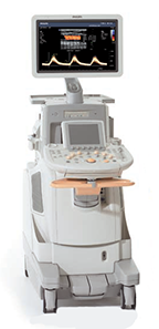
A revolution in premium performance ultrasoundThe Philips iU22 xMATRIX ultrasound system is premium performance ultrasound like you've never seen before.
Unique xMATRIX technology on the X6-1 PureWave transducer harnesses the power of over 9,000 active elements, more than 35 times greater than today's conventional transducers, to capture crisp, high-resolution images of even technically challenging patients.
Below is a generalized description of this systems technology, specifications, features and options. The below may not reflect the features and options available on units in our inventory. |
|
- Technologies
- Specifications
- Measurements & Reporting
- Transducers
- Tutorials
Pushing the boundaries
In today’s busy and demanding healthcare environment, you can rely on the iU22 system for superb image quality for all patient types, advanced volume imaging solutions, innovative workflow tools and exceptional scanning ease.
Exceptional 2D and 3D image quality from a single transducer |
|
| The new X6-1 PureWave xMATRIX transducer delivers outstanding 2D images, and then, with the touch of a button, converts to 3D to provide a more complete picture of anatomy without workflow disruption. | |
Real-time, simultaneous imaging in two planes |
|
| The X6-1 PureWave xMATRIX transducer features xPlane, which allows imaging in two planes simultaneously, without manually rotating the transducer. | |
Volume viewing on any PACS |
|
|
For the first time ever, you can send 3D MPRs to any PACS system, making volume images available wherever they are needed for decision-making, review and storage. |
|
Gain a better perspective of needle location during biopsies and ablations |
|
| The iU22 xMATRIX provides integrated image fusion and instrument navigation capabilities with PercuNav. The PercuNav system acts like a GPS for medical instruments when guiding soft tissue biopsy and ablation procedures. | |
Enhancements in breast imaging |
|
| The iU22 xMATRIX's strain-based breast elastography feature provides a highly sensitive exam tool for detecting breast anomalies. The Advanced Breast Tissue Specific Imaging (TSI) preset on the L12-5 transducer, helps users distinguish between cystic and solid masses in the breast. | |
SmartExam tools deliver efficiency |
|
| SmartExam guided workflow on the iU22 xMATRIX increases consistency and speed by automatically planning and processing application protocols. | |
xMATRIX |
|
| xMATRIX revolutionary ultra thin slice imaging delivers extraordinary tissue uniformity for improved textural pattern recognition, superb discrimination of small structures in the near, mid and far, and exceptional image quality so you can see anatomical details not seen with conventional ultrasound. | |
QLAB |
|
| QLAB provides automated and objective methods for quantifying ultrasound data to improve your workflow A full suite of plug-ins is available to customize QLAB's capabilities to suit your needs. | |
Elastography |
|
| Philips strain-based breast elastography technology is highly sensitive—obtained merely by patient breathing or cardiac motion. Elastography, on the iU22 xMATRIX with L12-5 transducer and Advanced Breast Tissue Specific Imaging (TSI) preset, enables you to differentiate relative stiffness of tissue through sonographic examination. | |
Digital Broadband Beamforming |
|
| Broadband beamforming uses the full range of ultrasound frequencies to capture the entire tissue signature information, preserving the quantity and quality of data through capture and preservation of the entire bandwidth of ultrasound signals. | |
SonoCT |
|
| SonoCT imaging technology uses transmit beam-steering techniques to obtain coplanar, tomographic images from different viewing angles, then combines these micro-angulated images into a single compounded image at real-time frame rates. | |
XRES |
|
| XRES adaptive image processing reduces artifacts and improves margin and border definition. | |
Volumetric Imaging |
|
| Capture volumes of data through remarkable visualization including iSlice, thick slice, invert, color invert, slice plane, and automated stacked contours. | |
PureWave |
|
| PureWave crystal technology improves penetration in difficult-to-image patients; revealing details of fine structures and increasing exam efficiency. | |
Tissue Aberration Correction |
|
| Tissue aberration correction technology corrects for speed of sound changes through adipose tissue, sharpens spatial resolution, reduces the effects of acoustic beam distortion, and improves tissue uniformity throughout the depth of view. |
System Specifications |
|
|
|
User Interface |
|
|
|
Imaging Modes |
|
|
|
Doppler Mode |
|
|
|
Software Technologies |
|
|
|
Connectivity Ports |
|
|
|
Image File Format |
|
|
|
Onboard / External Storage |
|
|
|
Power Supply |
|
|
|
Peripherals (Options) |
|
|
|
System Applications & Reporting |
|
|
|
Pain Management Imaging |
|
|
|
Women's Imaging |
|
|
|
Cardiac Imaging |
|
|
|
Small Parts |
|
|
|
Urology Imaging |
|
|
|
Vascular Imaging |
|
|
|
Structured Reporting |
|
|
User-Programmable Formulas & Tables |
Transducers / Probes (*Displayed MHz range includes multiple transducers) |
|
|
|
Cardiac Transducers/Probes |
|
|
|
3D / Volume Transducers/Probes |
|
|
|
Philips iU22 Ultrasound System User Guides - PDF |
|