Sonoline G40 Ultrasound
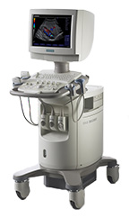
Comprehensive capabilities in a compact size.The SONOLINE G40™ ultrasound system brings
With a comprehensive range of features including color Doppler and pulsed-wave Doppler capabilities, the G40 system sets a new performance standard in the world of compact, mobile, color Doppler ultrasound systems.
Below is a generalized description of this systems technology, specifications, features and options. The below may not reflect the features and options available on units in our inventory. |
- Technology
- Specifications
- Measurements & Reporting
- Transducers
- Tutorials
The SONOLINE G40™ ultrasound system brings the benefits of Siemens core technology migration to an entirely new level of diagnostic performance and workflow efficiency, all in an ultra-portable system that meets your clinical needs. With a comprehensive range of features including color Doppler and pulsed-wave Doppler capabilities, the G40 system sets a new performance standard in the world of compact, mobile, color Doppler ultrasound systems.
- Migration of Siemens all-digital signal processing technology ensures best-in-class image quality for higher diagnostic confidence
- State-of-the-art beamforming architecture advances clinical capabilities with SynAps™ synthetic aperture technology for improved penetration and focus, parallel B-mode and color signal processing and phased array imaging functionality
- AutoColor technology provides single-step color Doppler optimization and autotrace functionality offers easy acquisition and quantification of color and Doppler studies
- Patented Hanafy lens acoustic technology enables clear differentiation of near-field structures and reduces far-field beam spread
- Imaging frequencies from 2–13 MHz provide superb support for a wide range of clinical applications
- Virtual Format Imaging increases anatomic information with a touch of a button, providing a selection of linear, trapezoidal and steered 2D imaging formats
- Open system architecture enables easy integration of advanced capabilities including phase inversion Tissue Harmonic Imaging (THI) and 3D imaging
- Comprehensive DIMAQ-IP integrated workstation allows instantaneous storage, review and quantification of complete ultrasound studies
- Compact, lightweight system with small footprint provides virtually effortless mobility and easy micropositioning in tight workspaces
- Intuitive user interface and familiar SONOLINE control panel layout advance workflow efficiency and clinical throughput
- Compact system design combined with color Doppler capability supports portable examinations without compromise
- TGO™ tissue grayscale optimization technology provides instantaneous, one-button image optimization
- Integrated documentation peripherals and onboard storage conveniently offer easy access to accessories needed to complete a
- comprehensive exam
- DIMAQ-IP integrated workstation supports capture and review of entire clinical studies in any environment, along with exportation of exams to CD
- Plug-and-play connectivity solutions allow easy integration into DICOM-enabled networks and PC-based workstations for efficient export of exam data
- Modern system design with integrated cable management and three transducer ports provides excellent protection during transport
System Specifications |
|
|
|
Monitor |
|
|
|
User Interface |
|
|
|
Operating / Display Modes |
|
|
|
Hard Drive |
|
|
|
CD-R/W Drive |
|
|
|
Power Supply |
|
|
|
Patient Study Management |
|
| Replay of digitally stored images in a selectable 1-up, 4-up, 9-up or 16-up screen format. The patient study screen allows for study selection, search, and deletion or for export to CD-R. | |
|
|
General Imaging |
|
|
|
Pain Management Imaging |
|
|
|
Women's Imaging |
|
|
|
Cardiac Imaging |
|
|
|
Small Parts |
|
|
|
Urology Imaging |
|
|
|
Vascular Imaging |
|
|
|
Transducers / Probes |
|
|
|
Cardiac Transducers/Probes |
|
|
|
Transducer Ports |
|
|
|
*Displayed MHz range includes multiple transducers